quinta-feira, 26 de fevereiro de 2009

Microvolt T-Wave Alternans and the MASTER Trial

Topic(s):  Arrhythmias
Date Posted: 1/27/2009

1. What is T-wave alternans?

T-wave alternans refers to a beat-to-beat alteration in the amplitude of the T-wave portion of the electrocardiogram tracing. These changes are small and are imperceptible to the naked eye.

2. What is microvolt T-wave alternans (MTWA) testing?

MTWA is a noninvasive technique used measure T-wave alternans, and is a risk stratification tool to predict the likelihood of fatal arrhythmias and sudden cardiac death in patients with a history of myocardial infarction or cardiomyopathy.

3. What is the clinical utility of TWA testing?

Prior trials have suggested that a negative T-wave alternans test indicates there is minimal risk of the patient developing ventricular tachyarrhythmias (VTA) and can therefore influence the treatment plan by sparing the risk and expense of surgery for implantation of an implantable cardioverter-defibrillator (ICD). Conversely, a positive test result will allow more aggressive treatment of patients at high risk for sudden cardiac death.

4. What was the purpose of the MASTER Trial?

The purpose of the MASTER trial was to evaluate use of MTWA testing for risk stratification for life-threatening ventricular tachyarrhythmic events among post-myocardial infarction (MI) patients with impaired ejection fraction undergoing ICD implantation. The hypothesis was that MTWA would predict ventricular tachyarrhythmias among patients with impaired ventricular function after MI.

5. What were the findings of the MASTER trial?

The MTWA test was non-negative in 63% of patients and negative in 37%. There was no significant difference in the annual rate of VTAs between patients with non-negative and negative tests (6.3% and 5.0%, respectively). MTWA was not a predictor of VTEs. Total mortality was twice as high in non-negative patients than in the patients with a negative MTWA test.

6. What was the conclusion of the MASTER Trial?

MTWA does not accurately identify post-infarction patients with ischemic cardiomyopathy who are at low risk of life-threatening arrhythmias.

7. What is the clinical significance of the trial?

The patients in this study met the MADIT-II criteria for ICD implantation (CAD, EF <>

Effects of Alcohol on Atrial Fibrillation: Myths and Truths

Carlos E.B. Balbão; Angelo A.V. de Paola; Guilherme Fenelon

Ther Adv Cardiovasc Dis.  2009;3(1):53-63.  ©2009 London: SAGE
Posted 02/17/2009

Abstract

Alcohol is the most consumed drug worldwide. Both acute and chronic alcohol use have been associated with cardiac arrhythmias, in particular atrial fibrillation, or so-called 'holiday heart syndrome'. Epidemiological, clinical and experimental studies have attempted to elucidate the mechanisms involved in this association. However, because most of these studies have shown conflicting results, the connection between ethanol and atrial arrhythmias remains controversial. Historical, epidemiological and pharmacological aspects of alcohol, as well as recent concepts on atrial fibrillation are reviewed. We then examine the literature and provide a critical point of view on the still elusive association between alcohol and atrial fibrillation.

Historic Background

The use of alcoholic beverages dates back to prehistoric times. Their medical use was recorded as early as the cuneiform writings in the Mesopotamia of 2200 BC. Out of the approximate 800 medications in ancient Egypt, some 15% included different kinds of beer or wine in their formulation. References to wine are also commonly found in the Old Testament. The Book of Genesis tells that Noah planted a vine after the Great Flood and got drunk. Interestingly, wine and beer could be mixed with other drugs. Thus, highly powerful beverages were produced at a time when distillation was still unknown.

The word alcohol comes from the Arabic word alkuhl, which means essence. Distillation techniques were developed by the Arabs during the Middle Ages, around 800 AD. Alchemists were fascinated by the invisible 'spirit' distilled from wine. They believed alcohol was the elixir of life and used it therapeutically. Worldwide, alcoholic beverages are consumed in multiple circumstances. Alcohol has been consumed for a long time in many different cultures, and consumption may vary within the same culture. As a consequence of such cultural diversity, attitudes towards alcohol have varied from tolerance to censure.

Epidemiological Aspects of Alcohol

Alcohol is currently the number one drug in the world in consumption levels. It has become a serious public health problem. Alcohol addiction affects from 10% to 12% of the population worldwide [WHO, 1999]. In the US, the National Household Survey on Drug Abuse published in 1996 showed that in the month previous to the research, 23.8% of men and 8.5% of women referred an episode of massive alcohol intake, which means five or more drinks at the same occasion at least once in that month. Heavy intake of ethanol is generally defined as the consumption of seven or more drinks per week by women, or over three doses at one time, and over fourteen drinks per week for men, or over four drinks at one time [US Department of Health and Human Service, 1995]. The financial burden from drug abuse is astonishing. Total annual economic cost resulting from the abuse of tobacco, alcohol and other drugs exceeds 238 billion dollars in the US. From that amount, alcohol alone accounts for 98.6 billion every year [Institute for Health Policy, 1993]. In the same period, at least 14 million Americans were reported as alcohol abusers or alcoholics. Those figures are most likely higher today and they reflect a world trend. It should be pointed out that clinical diagnosis and treatment are usually postponed to the point when the disease reaches an advanced stage and is already associated with a number of social and clinical complications that make treatment much more difficult.

Ethanol Pharmacology

Ethanol or ethyl alcohol (CH3CH2OH) is quickly absorbed in the stomach (20%) and in the small intestines (80%). After intake, maximum plasma concentration is reached between 30 and 90 min. As a result of ethanol first-pass metabolism through gastric and hepatic alcohol dehydrogenase (ADH), oral intake of ethanol results in lower blood levels when compared with IV administration. Following zero-order kinetics, which means being constant along time and independent of plasma concentrations, alcohol distribution over all tissues and body liquids is fast and uniform, and crosses the haematoencephalic and placental barriers. Redistribution velocity to specific tissues – as occurs with volatile anaesthetics – depends mostly on blood flow. Ethanol is highly liposoluble: 90% is metabolized in the liver, and from 5% to 10% is excreted without any change via exhaled air and urine. Ethanol oxidation occurs first into acetaldehyde and then into acetic acid. The intermediate metabolite acetaldehyde (CH3COH) is a reactive, toxic component and may be a contributor to hepatotoxicity.

Typically, alcohol content ranges from 4% to 6% in beer, 10–15% in wine and 40% or more in distilled beverages. The alcoholic degree on beverage labels corresponds to alcoholic percentage multiplied by two (e.g. 40% means that alcoholic degree is 80). Curiously enough, and as opposed to what lay individuals think, the size of alcoholic beverages containers is designed to contain approximately 14 g of alcohol per serving (which means to say 0.3 mol of ethanol), whether in a glass of beer (350 ml), in a glass of wine (150 ml) or in a glass of whisky or any other distilled beverage (45 ml). When consumed by a 70 kg individual, any of those beverages will result in a 30 mg% blood alcohol concentration. Alcohol consumption is measured per unit. One unit corresponds to 10 g of alcohol. In order to obtain the units equivalent to one given beverage, the amount taken must be multiplied by alcoholic concentration. That way, the absolute amount of alcohol in that beverage is calculated. Then, the following conversion is applied: one unit for every 10 g of alcohol contained in that beverage.

Clinical Effects of Ethanol

The acute clinical effects of ethanol are correlated to plasma concentration. When alcohol blood concentration reaches approximately 30 mg%, patients exhibit euphoria, excitement and slight changes in attention span; at 50 mg%, patients exhibit discreet motor discoordination and personality and behaviour changes; at 100 mg%, patients exhibit pronounced motor discoordination with ataxia, reduced concentration, worsening of sensitive reflexes and compromised humour; at 200 mg%, ataxia is aggravated and nausea and vomiting may occur; at 300 mg%, dysartria, amnesia, hypothermia and anaesthesia (stage I) may occur; at 400 mg%, coma and death can occur as a result of changes in central respiratory drive and/or systemic arterial hypotension. Blood levels of ethanol may vary depending on a number of factors; for instance, the time taken for alcohol intake, gender, body weight and water content in the body, as well as gastric emptying levels and the metabolism of individuals. On average, three conventional doses of alcohol (approximately 42 g of ethanol) while fasting leads to a maximum serum concentration ranging from 67 to 92 mg/dl in men. After a meal, the same dose will result in serum concentration ranging from 30 to 53 mg/dl. Serum concentrations are higher in women when compared with men when the same dose is considered. That may be explained by their lower average stature, their lower water content per body weight and their reduced gastric dehydrogenase activity when compared with men. Ethanol is metabolized at the speed of one conventional dose every 60–90 min in individuals who have normal hepatic function.

At low and moderate doses (one to three doses a day, ≤15 g/day for women and ≤30 g/day for men) ethanol has a protective effect in coronary cerebrovascular and peripheral vascular artery diseases and in metabolic syndrome, as demonstrated by large epidemiological studies such as the Framingham [Friedman and Kimball, 1986] and Physician's Health Study [Albert et al. 1999; Camargo et al. 1997], and also in a meta-analysis [Di Castelnuovo et al. 2002].

Ethanol has toxic effects on the myocardium, which are dose related and most prominent in long-term users [Patel et al. 1997; Preedy et al. 1994]. Fatty acids esters formed from ethanol enzyme reaction with free fatty acids and acetaldehyde seem to play a key role in the development of diffuse myocardial hypokinesia. This alcoholic cardiomyopathy is more prevalent among men and is characterized by cardiomegaly, diffuse hypokinesia and pathologic changes (dilation, fibrosis) in the myocardium of both ventricles. That applies to patients whose intake is at least 80 g of ethanol daily for a period of at least ten years. Electron microscopy studies have revealed ultrastructural myocardial damage including separation of filaments and loss of striation in addition to injuries in myofibril Z lines, with contractility loss at advanced stages of the disease, and dilation and rupture of mitochondrial crests. Other characteristics include lipid deposits (in particular triglycerides), fibrosis, widening of junctions, and damage to sarcoplasmic reticulum. At the final stages of the disease, inflammatory infiltrates can be observed. Acute alcoholic intoxication may also reduce myocardial contractility through direct effects of ethanol or acetaldehyde on troponin–tropomyosin coupling that is mediated by calcium inhibition [Horton and White, 1996; Guarnieri and Lakatta, 1990], and/or strong reduction of protein synthesis, particularly by acetaldehyde [Vary et al. 2005] and/or release of toxic free radicals to cardiac muscle [Atkinson et al. 1992].

Atrial Fibrillation Mechanisms

Atrial fibrillation is the most common sustained arrhythmia in clinical practice. Incidence increases with age, advancing from 0.5% in the sixth decade in life to approximately 10% of individuals over 80 years old. It is a significant risk factor for cerebral thromboembolism and is associated with an increased mortality [Abusaada et al. 2004; Friberg et al. 2003; Benjamin et al. 1994; Wolf et al. 1987, 1991, 1996]. The understanding of the mechanisms implicated in the genesis of atrial fibrillation has grown substantially in the last decade, which has led to the development of more effective clinical and interventional treatments, particularly catheter ablation techniques. In spite of this, its basic mechanisms have not been fully understood [Nattel and Opie, 2006]. Clinical and experimental evidence has suggested that atrial fibrillation patients may have atrial histological and/or electrophysiological changes, thus triggering the onset of arrhythmia as well as its perpetuation. However, in some situations, it may not be clear whether those changes are the cause of or the consequence from atrial fibrillation. Nevertheless, some cardiac and non-cardiac factors have been consistently associated with this kind of arrhythmia. Among the cardiac factors deserves mention any cause that leads to increases in left atrium dimensions (e.g. mitral valve disease, aortic valve disease, systemic arterial hypertension, pericarditis, myocarditis), left ventricular dysfunction, and the post-operative period following cardiac surgery. Among the non-cardiac factors: thyreotoxicosis, electrolyte disorders, drugs (both legal and illegal drugs), and alcohol abuse, especially acute alcohol abuse, but chronic as well.

Atrial tissue changes are involved in atrial fibrillation genesis, affecting the refractory periods and dispersion, as well as conduction velocity and triggering factors [West and Landa, 1962]. Solid evidence has been available to show that atrial fibrillation is based on multiple, continuous inter-atrial re-entries [Rensma et al. 1988; Moe and Abildskov, 1959], but recently the concept of focal atrial fibrillation has gained renewed interest [Nattel and Opie, 2006]. It has been consistently shown that pulmonary veins foci are the most relevant triggers of atrial fibrillation, but foci originating in the left atrial posterior wall, crista terminalis and caval veins have also been described. The intrinsic mechanisms of these foci remain uncertain, and rotors, anisotropic re-entry and automatic or triggered activity could play a role.

Given the major role of re-entry in the development and maintenance of atrial fibrillation, it is important to briefly review the conditions required to set up a re-entrant circuit. Re-entry requires that (1) the impulse blocks in a unidirectional fashion; and (2) the recirculation time of the impulse to the original site has to be longer than the refractory period of the proximal segment of the circuit. Should recirculation time be shorter, the impulse will reach the original site during refractoriness of the proximal segment of the circuit and re-entry will not be completed. In other words, the anatomic length of the circuit must be identical or longer than the distance travelled by the activation wave during the refractory period. This seminal concept defines the so-called wavelength, which corresponds to the product of the refractory period and conduction velocity [Wiener and Rosenblueth, 1946]. In the same tissue mass, shorter wavelengths – whether resulting from short refractory periods, slow conduction velocity, or both – are more bound to develop re-entry circuits than longer wavelengths. Wavelength is the major determining factor for atrial fibrillation inducibility via re-entry [Rensma et al. 1988].

Finally, in vivo monophasic action potential recordings provide additional key information for the electrophysiologic evaluation of cardiac arrhythmia mechanisms, since it keeps reasonable correlation with transmembrane action potential [Franz, 1991]. Thus, they are useful in determining refractory period changes. However, it is important to remark that the autonomic nervous system may cause major variations in the electrophysiologic parameters mentioned earlier. Therefore, accurate evaluation of those parameters should be undertaken in the presence and absence of complete autonomic blockade. These basic concepts are necessary for appreciation of the mechanisms by which alcohol could promote the genesis of atrial fibrillation.

Alcohol and Cardiac Arrhythmias

The association between alcohol use – whether acute or chronic – and cardiac arrhythmias has been widely described in the literature. In 1978, Ettinger et al. described a clinical syndrome they defined as 'acute changes in cardiac conduction or rhythm, associated to the ingestion of high amounts of alcohol in individuals with no other evidence of cardiac diseases, and which disappear without sequelae under abstinence'. As assistance to those patients was higher at certain week days (from Saturday to Tuesday), and on holiday season (between December 24 and January 1), the condition was named 'holiday heart syndrome'. The major electrocardiographic change found in that syndrome was the onset of supraventricular arrhythmias, particularly atrial fibrillation, which led the authors to infer that those patients had reduced atrial fibrillation threshold. The description was based on the observation of 24 alcoholics, with a total of 32 hospital admissions. The patients showed premature beats or tachyarrhythmias, especially atrial fibrillation. From those 32 hospital admissions, 19 occurred between Saturday and Tuesday. The remaining six episodes occurred between Christmas and New Year.

Other reports on acute atrial fibrillation and alcohol that are worth pointing out are as follows. Thornton et al. (1984) described four cases of acute atrial fibrillation and alcohol. Loewenstein et al. (1983) and Rich et al. (1985) considered alcohol as the cause for atrial fibrillation in 30–60% of patients presenting no cardiac condition, especially in those under 60 year old. Cohen et al. (1988) showed relative risk to be two-fold among those consuming high doses of ethanol (>6 doses/day) as compared to those consuming little ethanol (<1>et al. (1990, 1987) and Kupari and Koskinen (1991), while assessing ethylic populations through a questionnaire, observed that 42% of all cases of isolated atrial fibrillation among middle-aged men consuming over 150 g of ethanol/week was due to alcohol. Whyte et al. (2004) described one case of a freestyle skier who presented an atrial fibrillation episode during an exercise test. Later, the skier informed that he had ingested 12 units of alcohol on the previous day. After four weeks of abstinence, the test was repeated and no atrial fibrillation was observed. Finally, Koul et al. (2005) described one case of alcohol-induced atrial fibrillation in a 16-year-old male.

Controversies abound, starting with the term 'holiday heart syndrome'. Koskinen et al. (1987) and Kupari and Koskinen (1991) found correlation between ethanol consumption and atrial fibrillation. However, as opposed to the report by Ettinger et al. (1978), the incidence of that arrhythmia was higher on weekdays. Another conflicting aspect is whether the development of atrial fibrillation would occur during acute ingestion of alcohol, some hours later, or in the hangover period.

Epidemiological Studies

No correlation between ethanol ingestion and atrial fibrillation could be found by some major epidemiological studies, such as the one conducted in Framingham [Benjamin et al. 1994], the Manitoba study [Krahn et al. 1995], the Multifactor Primary Prevention study [Wilhelmsen et al. 2001], and the Renfrew/ Paisley study [Stewart, 2001]. The Cardiovascular Health study [Psaty et al. 1997] reported a reduced risk of atrial fibrillation depending on the alcohol dose ingested. A Danish study comprising 47,949 participants, in turn [Frost and Vestergaard, 2004], reported increased risk of atrial fibrillation or atrial flutter in males ingesting alcohol at least twice a week. The same authors could not, however, correlate alcoholic acute intoxication episodes to atrial fibrillation episodes in women, which was explained by the lower consumption of ethanol by females. In an analysis of the Framingham study [Djousse et al. 2004], comparing alcohol ingestion and counting on a control group, the follow-up of 10,333 patients for a longer-than-50-year period, with 1,055 cases of atrial fibrillation in that time span (544 males and 511 females), the association between moderate alcohol consumption and atrial fibrillation was low, but significant among individuals ingesting over 36 g/day of ethanol, which means to say more than 3 drinks per day (34% increased risk of AF – 95% CI 1–78%). Planas et al. (2006) investigated 115 patients reporting their first atrial fibrillation episode in Catalonia, Spain. In the six-month follow-up, 32 patients (27.8%) reported relapses. The authors concluded that the risk of idiopathic atrial fibrillation recurrence was high, and was enhanced by moderate alcohol consumption and increased left ventricular ectopic activity, probably of sympathetic origin. This trend was less marked in paroxysmal atrial fibrillation of vagal origin. In the Copenhagen City Heart study, Mukamal et al. (2005) described increased risk of atrial fibrillation (RR 1.45, 95% CI 1.02– 2.04) in males consuming more than 35 drinks per week. While assessing 1,232 atrial fibrillation cases in the Cardiovascular Health study on an average follow-up time of 9.1 years, Mukamal et al. (2007) also concluded that current moderate alcohol consumption is not associated with the risk of atrial fibrillation or with risk of death after diagnosis of atrial fibrillation, but former drinking identifies individuals at higher risk. Guize et al. (2007) studied atrial fibrillation prevalence in France. In a large population (98,961 males and 55,109 females), with average follow-up time of 15.2 years, alcohol consumption showed to be associated with atrial fibrillation only in men [OR = 1.7 (1.2–4)]. In a prospective study with a control group, Marcus et al. (2008) recently evaluated 195 consecutive patients that had been referred for atrial fibrillation ablation or atrial flutter in a period of two years. A significant, positive association between alcohol use and atrial flutter was reported in younger patients. The authors inferred that a possible mechanism for alcohol action could have been linked to high right atrium effective refractory period reduction in those patients.

Despite these controversial results, it seems reasonable to conclude that chronic overconsumption of ethanol is a common risk factor for atrial fibrillation in an otherwise healthy individual ( Table 1 ).

Electrophysiological Effects of Ethanol in Humans

Several studies with limited number of patients have been conducted attempting to elucidate how acute alcoholic promotes atrial arrhythmias, particularly atrial fibrillation. Greenspon and Schaal (1983) investigated 14 patients after ethanol infusion. They demonstrated that ethanol increased heart rate and reduced corrected sinus node recovery time. However, noteworthy is the fact that it did not change refractory periods. Gould et al. (1978) also investigated 14 patients. They observed that after alcohol infusion only the ventricular refractory period was shortened, as opposed to Engel and Luck (1983), who did not detect any change in atrial effective refractory period in the eleven patients investigated. The latter authors suggested that alcohol-related atrial arrhythmias might be attributed to intra-myocardial catecholamine release or to toxic direct effect of the metabolite acetaldehyde. Steinbigler et al. (2003) investigated 40 patients with a history of atrial fibrillation related to alcohol consumption with signal averaged ECG and demonstrated that those individuals had their P wave duration significantly prolonged by ethanol – a predisposing factor for atrial fibrillation. Maki et al. (1998) studied heart rate variability in six male patients with a previous history of ethanol-induced atrial fibrillation. The authors demonstrated that acute alcoholic intoxication leads to an increased sympathetic drive. Another pending issue is whether alcohol could cause arrhythmia upon withdrawal. This could be associated with high adrenergic responses and/or electrolyte disorders, particularly low potassium and magnesium levels. However, other authors, as Buckingham et al. (1985), Gribaldo et al. (1985) and Denison et al. (1994) have not observed an increased incidence of atrial or ventricular arrhythmias in the period through electrocardiographic monitoring.

In regard to electrocardiographic parameters in the literature, the few occurrences on ECG changes and alcohol refer to the chronic use of the drug. Uyarel et al. (2005) studied ten young, healthy volunteers and the changes in ECG after oral consumption of ethanol. They observed P wave duration increase. Lorsheyd et al. (2005) also studied ten healthy, young volunteers. After acute ethanol intake, the authors found PR and QTc interval increase. Those studies were conducted with a small number of patients who had an intact autonomic nervous system. Only surface ECG was evaluated. No other electrophysiologic parameters were investigated.

A few studies [Engel and Luck, 1983; Greenspon and Schaal, 1983] evaluating the effects of ethanol on inducibility of atrial arrhythmias were undertaken in alcoholics, and suggested that chronic alcohol ingestion may increase vulnerability to atrial arrhythmias. However, whether these findings are applicable to nonalcoholic subjects with normal hearts remain uncertain. In fact, one of the drawbacks in some of the above-mentioned clinical studies is that the patients themselves, through a questionnaire, provided the information on alcohol consumption. The most commonly used questionnaire was CAGE, with 81–91% sensitivity, and 77– 89% specificity for the discrimination of alcoholic and non-alcoholic patients.

Based on previous studies, it is fair to state that chronic consumption of alcohol may create a substrate that eventually increases vulnerability to atrial arrhythmias ( Table 1 ). However, the mechanisms responsible for this proarrhythmic response remain obscure.

Electrophysiological Effects of Ethanol in Experimental Studies

Most studies conducted to evaluate the electrophysiologic properties of alcohol were based on in vitro isolated preparations of heart cells. However, notwithstanding the relevance of these studies, one has to keep in mind the limitations of this kind of preparations, such as the high concentrations of ethanol used in the perfusate, when compared with in vivo models. Thus, in vitro findings should not be directly extrapolated to the clinical arena. Some of the in vitro studies that should be mentioned include the study by Williams et al. (1980), using isolated canine and swine cells. The authors demonstrated a reduction (around 8%) of transmembrane action potential duration and inferred that such alteration was secondary to decreased calcium currents. The study by Habuchi et al. (1995) using ventricular cells of swine demonstrated that the observed calcium channel inhibition – both under acute and chronic alcoholic intoxication – was responsible for the negative inotropic effect through action potential reduction and the development of arrhythmia. The study conducted by Snoy et al. (1980) showed the same results. Carpentier and Gallardo-Carpentier (1987) used rat cells, and demonstrated increased automaticity of sinoatrial cells. Opposite results also related to the automaticity of rat atrial cells were reported by Jain and Carpentier (1998). Of note, while studying cardiomyocytes from the pulmonary veins of rabbits and counting on a control group (with no perfusion with ethanol), Chen et al. (2004, 2002, 2001) demonstrated that although alcohol reduces action potential duration, it did not increase the incidence of delayed afterdepolarizations of pulmonary veins cardiomyocytes, in contrast to what had been shown by the same group during the perfusion of the same cells with thyroid hormone or after fast stimulation. This finding suggests that ethanol has no direct effects on the arrhythmogenic potential of these cells.

Although in vitro studies suggest that alcohol at high concentrations has a depressant effect on calcium currents ultimately decreasing action potential duration, these observations have not been reproduced in intact animal models ( Table 1 ). From the in vivo experimental studies, those worth mentioning are the following: in an experimental swine model, Anadon et al. (1996) demonstrated that high doses of alcohol (higher than those tolerated by humans) facilitated induction of atrial fibrillation and atrial flutter, but evaluation of atrial electrophysiological parameters and experiments in sham controls were not performed. In a canine model, Goodking et al. (1975) observed that alcohol had a depressant effect in atrioventricular conduction, but did not alter intraventricular conduction. However, the authors did not investigate the autonomic influences and had no sham control group. Kostis et al. (1977), also using dogs, reported that paradoxically to what had been previously demonstrated, alcohol showed an antiarrhythmic effect, and that the atrial fibrillation very often considered resulting from alcohol ingestion could have been due to electrolyte, autonomic or histological changes. Using canine models, Nguyen et al. (1987) observed that alcohol caused vasodilation, negative inotropic effect, and atrial antiarrhythmic effect. Madan and Gupta (1967) also observed alcohol atrial and ventricular antiarrhythmic effect in canine models.

Given the conflicting results and limitations of the previously mentioned investigations, we [Fenelon et al. 2007] have recently conducted a comprehensive study on the in vivo electrophysiological effects of acute alcoholic intoxication. We evaluated the cardiac electrophysiologic effects of ethanol in 23 anaesthetized dogs, with structurally normal hearts, at baseline and after two cumulative IV doses of alcohol or saline in the control group: first dose – 1.5 ml/kg (mean plasma level at 200 mg/dl); second dose – 1.0 ml/kg (279 mg/dl). Those doses correspond to acutely moderate and severe alcoholic intoxication, respectively. The dogs were divided into five groups: group I – ethanol group: closed chest, absence of autonomic blockade (n = 5); group II – sham control group (n = 3): closed chest, saline infused, rather than ethanol; group III – ethanol group: closed chest under complete pharmacological (atropine + propranolol) autonomic blockade (n = 5); group IV – ethanol group: closed chest, no autonomic blockade, for evaluation of left ventricular ejection fraction using 2D echocardiogram and biopsies of atrial tissue for histological and ultrastructural analysis (n = 5); and group V – ethanol group: absence of autonomic blockade, open chest, and biatrial epicardial mapping (n = 5). Haemodynamic, electrocardiographic and electrophysiologic parameters were assessed. In groups I, II and III, high right atrium monophasic action potential (MAP) recordings, measured at 90% of repolarization, were obtained with standard techniques [Franz, 1991] as previously reported [Fenelon and Brugada, 1998]. Group IV was evaluated for left ventricular function and atrial tissue was obtained in the same group for optical and electron microscopy. In group V, the chest was opened and an eight-bipole plaque was placed in Bachmann's bundle to measure interatrial conduction time, conduction velocity, and wavelength. As mentioned earlier, the wavelength is the best predictive parameter for the induction of re-entrant atrial arrhythmias [Rensma et al. 1988]. Inducibility of atrial arrhythmias was assessed with up to four extra stimuli and rapid burst pacing for 15 s duration. Atrial and ventricular tachyarrhythmias longer than 30 s in duration were considered sustained.

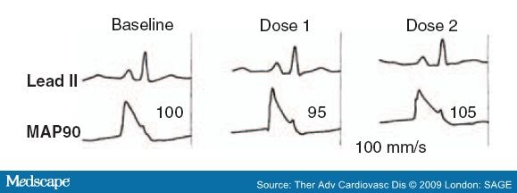
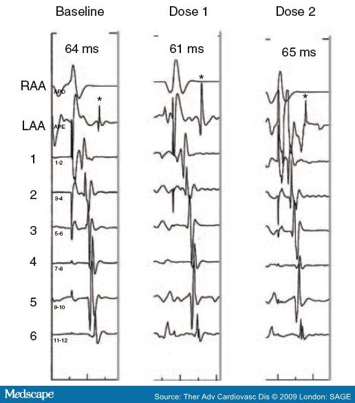
In groups I, II and III, ethanol was not shown to significantly alter hemodynamic variables (systolic, diastolic and mean blood pressure), electrocardiographic variables (P wave duration, QRS duration; PR and QT interval), or electrophysiologic parameters (Figure 1) (PA interval duration, HV interval, MAP duration, right atrium effective refractory period, corrected sinus node recovery time). In group V (open chest and biatrial epicardial mapping), ethanol did not affect interatrial conduction time (Figure 2), conduction velocity or wavelength. In all groups, no atrial or ventricular arrhythmias were induced in any dog. Histological and ultrastructural analysis was normal in all animals in group IV.

Figure 1. 

Electrocardiographic and right atrial monophasic action potentials (MAP) in ethanol-treated dogs without autonomic blockade. From top to bottom: ECG lead II and right atrial MAP. The duration of MAP at 90% repolarization (MAP90) in ms is indicated. Tracings recorded before (baseline) and after the first (dose 1) and second (dose 2) doses of ethanol. No appreciable changes occur after alcohol. Paper speed at 100 mm/s. Reproduced with permission from Fenelon G et al. (2007) Alcohol Clin Exp Res 31(9): 1574–1580.


Figure 2. 

Epicardial mapping in open-chest dogs is shown. Interatrial conduction during continuous right atrial appendage (RAA) pacing (200 ms cycle length) recorded before (baseline) and after the first (dose 1) and second (dose 2) doses of ethanol is depicted. Plaque bipole numbers and inter-atrial conduction time in milliseconds are indicated. Asterisks differentiate local left atrial appendage (LAA) activation from far field stimulus artifact. No appreciable changes in inter-atrial impulse propagation occur after alcohol. Paper speed at 200 mm/s. Reproduced with permission from Fenelon G et al. (2007) Alcohol Clin Exp Res 31(9): 1574–1580.


Ejection fraction was also evaluated in group IV and showed a significant reduction (77% vs 73% vs 66%; p = 0.04) with cumulative ethanol doses. Based on those results, and stressing the fact that it is an experimental canine model with normal hearts, moderate and high doses of alcohol promoted discreet, progressive left ventricular systolic dysfunction, did not alter electrocardiographic or electrophysiologic cardiac parameters, did not induce histological or ultrastructural changes in atrial tissue, and did not promote atrial or ventricular arrhythmia inducibility. These findings suggest that acute alcoholic intoxication does not exert direct myocardial actions that may create a substrate for the development of arrhythmias. The authors are not aware of any other study that has evaluated all those parameters concurrently, whether experimentally or clinically.

Conclusion

The 'holiday heart syndrome' was defined in 1978 as the occurrence of alcohol-induced arrhythmias, chiefly atrial fibrillation, in otherwise healthy individuals. Such arrhythmias may occur during acute alcohol intake and withdrawal. However, as mentioned earlier in this review, the association between alcohol ingestion and atrial fibrillation has been challenged by several epidemiological studies and remains controversial.

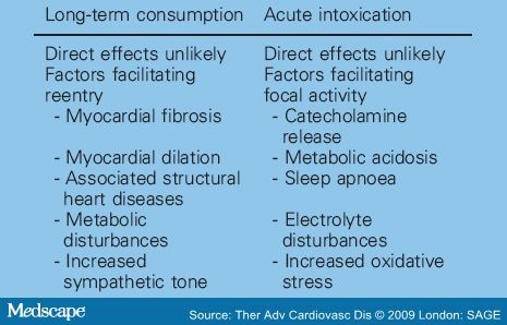
Our own and others' data suggest that acute alcohol ingestion, per se, does not render the atrium susceptible to atrial fibrillation. The observation that ethanol does not exert significant effects on atrial electrophysiology (refractory period, conduction velocity and wavelength) and arrhythmia inducibility supports the hypothesis that the development of atrial arrhythmias in the setting of alcoholic intoxication may require additional pathological conditions, such as metabolic disturbances, autonomic imbalance or sleep apnoea [Wong, 1973; Rosenqvist, 1998]. Corroborating this premise, although it is well known that ethanol directly affect myocardial contractile function, indirect effects on the myocardium may also occur. Both ethanol and its metabolite acetaldehyde have been shown to increase levels of circulating catecholamines [Williams et al. 1980]. Further, ethanol may also induce oxidative stress and release of plasma free fatty acids [Rosenqvist, 1998]. These prominent indirect effects of ethanol may be arrhythmogenic, particularly in individuals prone to atrial fibrillation such as patients with focal atrial fibrillation. Furthermore, other patients susceptible to atrial fibrillation include those with structural heart diseases and, possibly, long-term alcohol users in whom subclinical cardiac abnormalities may occur ( Table 2 ).

There is clearly a need for further studies evaluating the relationship between alcohol and atrial fibrillation in otherwise healthy individuals. However, these studies should contemplate the recent knowledge gained on the mechanisms of atrial fibrillation genesis, foremost focal atrial fibrillation. Further, the electrophysiological and structural effects of chronic alcohol consumption should be better characterized. These studies are key to determine if and how alcohol promotes atrial fibrillation.


Table 1. Summary of Studies Evaluating Alcohol and Atrial Fibrillation


Table 1: Summary of Studies Evaluating Alcohol and Atrial Fibrillation


    Table 2. Possible Mechanisms of Alcohol-induced Atrial Fibrillation


    Table 2: Possible Mechanisms of Alcohol-induced Atrial Fibrillation

      References

      1. Abusaada, K., Sharma, S.B., Jaladi, R. and Ezekowitz, M.D. (2004) Epidemiology and management of new-onset atrial fibrillation. Am J Manag Care 10: S50-57.
      2. Albert, C.M., Manson, J.E., Cook, N.R., Ajani, U.A., Gaziano, J.M. and Hennekens, C.H. (1999) Moderate alcohol consumption and the risk of sudden cardiac death among US male physicians. Circulation 100: 944-950.
      3. Anadon, M.J., Almendral, J., González, P., Zaballos, M., Delcan, J.L. and De Guevara, J.L. (1996) Alcohol concentration determines the type of atrial arrhythmia induced in a porcine model of acute alcoholic intoxication. Pacing Clin Electrophysiol 19: 1962-1967.
      4. Atkinson, L., Why, H., Ward, R.J., Peters, T.J. and Richardson, P.J. (1992) Antioxidant status in dilated cardiomyopathy and alcohol heart muscle disease. Alcohol Alcohol 28: 67-73.
      5. Benjamin, E.J., Levy, D., Vaziri, S.M., D'Agostino, R.B., Belanger, A.J. and Wolf, P.A. (1994) Independent risk factors for atrial fibrillation in a population-based cohort: The Framingham Heart Study. JAMA 271: 840-844.[Abstract]
      6. Buckingham, T.A., Kennedy, H.L., Goenjian, A.K., Vasilomanolakis, E.C., Shriver, K.K., Sprague, M.K. et al. (1985) Cardiac arrhythmias in a population admitted to an alcohol detoxification center. Am Heart J 110: 961-965.
      7. Camargo Jr, C.A., Hennekens, C.H., Gaziano, J.M., Glynn, R.J., Manson, J.E. and Stampfer, M.J. (1997) Prospective study of moderate alcohol consumption in US male physicians. Arch Intern Med 157: 79-85.
      8. Carpentier, R.G. and Gallardo-Carpentier, A. (1987) Acute and chronic effects of ethanol on sinoatrial electrophysiology in the rat heart. J Cardiovasc Pharmacol 10: 616-621.
      9. Chen, Y.C., Chen, S.A., Chen, Y.J., Tai, C.T., Chan, P. and Lin, C.I. (2004) Effect of ethanol on the electrophysiological characteristics of pulmonary vein cardiomyocytes. Eur J Pharmacol 483: 215-222.
      10. Chen, Y.J., Chen, S.A., Chen, Y.C., Yeh, H.I., Chan, P., Chang, M.S. et al. (2001) Effects of rapid pacing on the arrhythmogenic activity of single cardiomyocytes from pulmonary veins: implications in initiation of atrial fibrillation. Circulation 104: 2849-2854.
      11. Chen, Y.J., Chen, S.A., Chen, Y.C., Yeh, H.I., Chan, P., Chang, M.S. et al. (2002) Effects of thyroid hormone on the arrhythmogenic activity of pulmonary vein cardiomyocytes. J Am Coll Cardiol 39: 366-372.
      12. Cohen, E.J., Klatsky, A.L. and Armstrong, M.A. (1988) Alcohol use and supraventricular arrhythmia. Am J Cardiol 62: 971-973.
      13. Denison, H., Jern, S., Jagenburg, R., Wendestam, C. and Wallerstedt, S. (1994) Influence of increased adrenergic activity and magnesium depletion on cardiac rhythm in alcohol withdrawal. Br Heart J 72: 554-560.
      14. Di Castelnuovo, A., Rotondo, S., Iacoviello, L., Donati, M.B. and De Gaetano, G. (2002) Meta-analysis of wine and beer consumption in relation to vascular risk. Circulation 105: 2836-2844.
      15. Djousse, L., Levy, D., Benjamin, E.J., Blease, S.J., Russ, A., Larson, M.G. et al. (2004) Long-term alcohol consumption and the risk of atrial fibrillation in the Framingham study. Am J Cardiol 93: 710-713.
      16. Engel, T.R. and Luck, J.C. (1983) Effect of whiskey on atrial vulnerability and `holiday heart'. J Am Coll Cardiol 1: 816-818.[Abstract]
      17. Ettinger, P.O., Wu Chia, F., De La Cruz Jr, C., Weisse, A.B., Ahmed, S.S. and Reagan, T.J. (1978) Arrhythmias and the `Holiday Heart': alcohol-associated cardiac rhythm disorders. Am Heart J 95: 555-562.
      18. Fenelon, G. and Brugada, P. (1998) Unipolar waveforms and monophasic action potentials in the characterization of slow conduction in human atrial flutter. Pacing Clin Electrophysiol 21: 2580-2587.
      19. Fenelon, G., Balbao, C.E.B., Fernandes, R., Arfelli, A., Landim, P., Ayres, O. et al. (2007) Characterization of the acute cardiac electrophysiologic effects of ethanol in dogs. Alcohol Clin Exp Res 31: 1574-1580.
      20. Franz, M.R. (1991) Method and theory of monophasic action potential recording. Prog Cardiovasc Dis 33: 347-68.
      21. Friberg, J., Scharling, H., Gadsboll, N. and Jensen, G.B. (2003) Sex-specific increase in the prevalence of atrial fibrillation (The Copenhagen City Heart Study). Am J Cardiol 92: 1419-1423.
      22. Friedman, L.A. and Kimball, A.W. (1986) Coronary heart disease mortality and alcohol consumption in Framingham. Am J Epidemiol 124: 481-489.
      23. Frost, L. and Vestergaard, P. (2004) Alcohol and risk of atrial fibrillation or flutter - a cohort study. Arch Intern Med 164: 1993-1998.
      24. Goodking, M.J., Gerber Jr, N.H., Mellen, J.R. and Kostis, J.B. (1975) Altered intracardiac conduction after acute administration of ethanol in the dog. J Pharmacol Exp Ther 194: 633-638.
      25. Gould, L., Reddy, C.V., Becker, W., Oh, K.C. and Kim, S.G. (1978) Electrophysiologic properties of alcohol in man. J Electrocardiol 11: 219-226.
      26. Greenspon, A.J. and Schaal, S.F. (1983) The `holiday heart': electrophysiologic studies of alcohol effects in alcoholics. Ann Intern Med 98: 135-139.
      27. Gribaldo, R.G., Pomoni, G.S. and Sale, F. (1985) Arrhythmias and left function in chronic alcoholics with cirrhosis. Am J Cardiol 56: 825-827.
      28. Guarnieri, T. and Lakatta, E.G. (1990) Mechanism of myocardial contractile depression by clinical concentrations of ethanol. J Clin Invest 85: 1462-1467.
      29. Guize, L., Thomas, F., Bean, K., Benetos, A. and Pannier, B. (2007) Fibrillation atriale: prevalence, facteurs de risqué et mortalité dans une vaste population française suivie 15 ans. Bull Acad Natl Med 191: 791-803.
      30. Habuchi, Y., Furukawa, T., Tanaka, H., Lu, L.L., Morikawa, J. and Yoshimura, M. (1995) Ethanol inhibition of Ca2+ and Na+ currents in the guinea-pig heart. Eur J Pharmacol 292: 143-149.
      31. Horton, J.W. and White, J.D. (1996) Cardiac contractile and sarcoplasmic reticulum function after acute ethanol consumption. J Surg Res 64: 132-138.
      32. Institute for Health Policy, Brandeis University. (1993) Substance Abuse: The Nation's Number One Health Problem. Princeton, NJ: Robert Wood Johnson Foundation.
      33. Jain, A.K. and Carpentier, R.G. (1998) Cardiac electrophysiological actions and interactions of ethanol, cocaine, and the metabolite ethylcocaine. J Electrocardiol 31: 293-302.
      34. Koskinen, P., Kupari, M., Leinonon, H. and Luomanmaöki, K. (1987) Alcohol and new onset atrial fibrillation: a case - control study of a current series. Br Heart J 57: 468-473.
      35. Koskinen, P., Kupari, M. and Leinonen, H. (1990) Role of alcohol in recurrences of atrial fibrillation in persons less than 65 years of age. Am J Cardiol 66: 954-958.
      36. Kostis, J.B., Goodkind, J., Skvaza, H., Gerber Jr, N.H. and Kuo, P.T. (1977) Effect of alcohol on the atrial fibrillation threshold in dogs. Angiology J Vasc Dis 28: 583-587.
      37. Koul, P.B., Sussmane, J.B., Cunill-De Sautu, B. and Minarik, M. (2005) Atrial fibrillation associated with alcohol ingestion in adolescence: holiday heart in pediatrics. Pediatr Emerg Care 21: 38-39.
      38. Krahn, A.D., Manfreda, J., Tate, R.B., Mathewson, F.A.L. and Cuddy, T.E. (1995) The natural history of atrial fibrillation: incidence, risk factors, and prognosis in the Manitoba Follow-Up Study. Am J Med 98: 476-484.
      39. Kupari, M. and Koskinen, P. (1991) Time of onset of supraventricular tachyarrhythmia in relation to alcohol consumption. Am J Cardiol 67: 718-722.
      40. Lorsheyd, A., de Lange, D.W., Hijmering, M.L., Cramer, M.J. and van de Wiel, A. (2005) PR and QtC interval prolongation on the electrocardiogram after binge drinking in health individuals. Neth J Med 63: 59-63.
      41. Lowenstein, S.R., Gabow, P., Cramer, J., Oliva, P.B. and Ratner, K. (1983) The role of alcohol in new-onset atrial fibrillation. Arch Intern Med 143: 1882-1885.
      42. Madan, B.R. and Gupta, R.S. (1967) Effect of ethanol in experimental auricular and ventricular arrhythmias. Jpn J Pharmacol 17: 683-684.
      43. Maöki, T., Toivonen, L., Koskinen, P., Naöveri, H., Haörkonen, M. and Leinonen, H. (1998) Effect of ethanol drinking, hangover, and exercise on adrenergic activity and heart rate variability in patients with a history of alcohol-induced atrial fibrillation. Am J Cardiol 82: 317-322.
      44. Marcus, G.M., Smith, L.M., Whiteman, D., Tseng, Z.H., Badhwar, N., Lee, B.K. et al. (2008) Alcohol intake is significantly associated with atrial flutter in patients under 60 years of age and a shorter right atrial effective refractory period. Pacing Clin Electrophysiol 31: 266-272.
      45. Moe, G.K. and Abildskov, J.A. (1959) Atrial fibrillation as a self-sustaining arrhythmia independent of focal discharge. Am Heart J 58: 59-70.
      46. Mukamal, K.J., Psaty, B.M., Rautaharju, P.M., Furberg, C.D., Kuller, L.H., Mittleman, M.A. et al. (2007) Alcohol consumption and risk and prognosis of atrial fibrillation among older adults: the Cardiovascular Health Study. Am Heart J 153: 260-266.
      47. Mukamal, K.J., Tolstrup, J.S., Friberg, J., Jensen, G. and Gronbaek, M. (2005) Alcohol consumption and risk of atrial fibrillation in men and women - the Copenhagen City Heart Study. Circulation 112: 1736-1742.
      48. Nattel, S. and Opie, L.H. (2006) Controversies in atrial fibrillation. Lancet 367: 262-272.
      49. Nguyen, T.N., Friedman, H.S. and Mokraoui, A.M. (1987) Effects of alcohol on experimental atrial fibrillation. Alcohol Clin Exp Res 11: 472-476.
      50. Patel, V.C., Why, H.J., Richardson, P.J. and Preedy, V.R. (1997) The effects of alcohol on the heart. Adverse Drug React Toxicol Rev 16: 15-43.
      51. Planas, F., Romero-Menor, C., Vasquez-Oliva, G., Poblet, T. and Navarro-López, F. (2006) Historia natural y factores de riesgo de recurrencia de la fibrilación auricular primaria (Registro FAP). Rev Esp Cardiol 59: 1106-1112.
      52. Preedy, V.R., Siddiq, T., Why, H. and Richardson, P.J. (1994) The deleterious effects of alcohol on the heart: involvement of protein turnover. Alcohol Alcohol 29: 141-147.
      53. Psaty, B.M., Manolio, T.A., Kuller, L.H., Kronmal, R.A., Cushman, M., Fried, L.P. et al. (1997) Incidence of and risk factors for atrial fibrillation in older adults. Circulation 96: 2455-2461.
      54. Rensma, P.L., Allessie, M.A., Lammers, W.J.E.P., Bonke, F.I.M. and Schalij, M.J. (1988) Length of excitation wave and susceptibility to reentrant atrial arrhythmias in normal conscious dogs. Circ Res 62: 395-410.
      55. Rich, E.C., Siebold, C. and Campion, B. (1985) Alcohol-related acute atrial fibrillation, a case-control study and review of 40 patients. Arch Intern Med 145: 830-833.
      56. Rosenqvist, M. (1998) Alcohol and cardiac arrhythmias. Alcohol Clin Exp Res 22: 318S-322S.
      57. Snoy, F.J., Harker, R.J., Thies, W. and Greenspan, K. (1980) Ethanol-induced electrophysiological alterations in canine Purkinje fibers. J Stud Alcohol 41: 1023-1030.
      58. Steinbigler, P., Haberl, R., Koönig, B. and Steinbeck, G. (2003) P-wave signal averaging identifies patients prone to alcohol-induced paroxysmal atrial fibrillation. Am J Cardiol 91: 491-494.
      59. Stewart, S., Hart, C.L., Hole, D.J. and McMurray, J.J. (2001) Population prevalence, incidence, and predictors of atrial fibrillation in the Renfrew/Paisley study. Heart 86: 516-521.
      60. Thornton, J.R. (1984) Atrial fibrillation in healthy non-alcoholic people after an alcoholic binge. Lancet 2: 1013-1015.
      61. US Department of Health and Human Service. ( 1995) The Physician's Guide to Helping Patients with Alcohol Problems. Washington, DC: US Department of Health and Human Services, NIH Publication 95-3769.
      62. US Department of Health and Human Service ( 1996) Preliminary Estimates from 1995. National Household Survey and Drug Abuse. Advance Report 18., Rockville MD: National Institute on Drug Abuse.
      63. Uyarel, H.O.C., Karabulut, A., Okmen, E. and Cam, N. (2005) Acute alcohol intake and P-wave dispersion in health men. Anadolu Kardiyol Derg 5: 289-293.
      64. Vary, T.C., Deiter, G. and Goodman, A.S. (2005) Acute alcohol intoxication enhances myocardial eIF4G phosphorylation despite reducing mTOR signaling. Am J Physiol Heart Circ Physiol 288: H121-128.
      65. West, T.C. and Landa, J.F. (1962) Minimal mass required for induction of a sustained arrhythmia in isolated atrial segments. Am J Physiol 202: 232-236.
      66. Whyte, G., Stephens, N., Sharma, S., Shave, R., Budgett, R. and McKenna, W.J. (2004) Spontaneous atrial fibrillation in a freestyle skier. Br J Sports Med 38: 230-232.
      67. Wiener, N. and Rosenblueth, A. (1946) The mathematical formulation of the problem of conduction of impulses in a network of connected excitable elements, specifically in cardiac muscle. Arch Inst Cardiol Mex 16: 205-265.
      68. Wilhelmsen, L., Rosengren, A. and Lappas, G. (2001) Hospitalizations for atrial fibrillation in the general male population: morbidity and risk factors. J Intern Med 250: 382-389.
      69. Williams, E.S., Mirro, M.J. and Bailey, J.C. (1980) Electrophysiological effects of ethanol, acetaldehyde, and acetate on cardiac tissues from dog and guinea pig. Circ Res 47: 473-478.
      70. Wolf, P.A., Abbott, R.D. and Kannel, W.B. (1987) Atrial fibrillation: a major contributor to stroke in the elderly: The Framingham Study. Arch Intern Med 147: 1561-1564.
      71. Wolf, P.A., Abbott, R.D. and Kannel, W.B. (1991) Atrial fibrillation as an independent risk factor for stroke: The Framingham Study. Stroke 22: 983-988.
      72. Wolf, P.A., Benjamin, E.J., Belanger, A.J., Kannel, W.B., Levy, D. and D'Agostino, R.B. (1996) Secular trends in the prevalence of atrial fibrillation: The Framingham Study. Am Heart J 131: 790-795.
      73. Wong, M. (1973) Depression of cardiac performance by ethanol unmasked during autonomic blockade. Am Heart J 86: 508-515.
      74. World Health Organization (WHO) (1999) Global Status Report on Alcohol: 1999. Geneva: WHO. Available at: http://www.who.int/substance_abuse/publications/en/GlobalAlcohol_overview.pdf

      terça-feira, 24 de fevereiro de 2009

      Outcomes in Adolescents with Postural Orthostatic Tachycardia Syndrome Treated with Midodrine and [beta]-Blockers.

      LAI, CINDY, FISCHER, PHILIP, BRANDS, CHAD, FISHER, JENNIFER, PORTER, CO-BURN, DRISCOLL, SHERILYN, GRANER, KEVIN

      Pacing & Clinical Electrophysiology. 32(2):234-238, February 2009.

      Background: Postural orthostatic tachycardia syndrome (POTS) is associated with debilitating fatigue, dizziness, and discomfort in previously healthy adolescents. The effects of medical therapy have not been well studied in this patient population. This study assessed the relative efficacy and impact of drug therapy on the functioning and quality of life in adolescents with POTS.

      Methods: A retrospective, single center, chart review analysis with a follow-up written survey was conducted on a group of 121 adolescents who had undergone autonomic reflex screening at the Mayo Clinic from 2002 to 2005 as part of an evaluation for possible POTS.

      Results: Of 121 surveys sent, 47 adolescents returned a completed survey. In this cohort of patients, the two most commonly prescribed drug therapies were midodrine (n = 13) and [beta]-blockers (n = 14). Patients in the midodrine group were comparable to patients in the [beta]-blocker group in gender, age, pretreatment postural heart rate changes, and months from initial evaluation to survey completion. More patients treated with a [beta]-blocker reported improvement after visiting Mayo Clinic (100% vs 62%, P = 0.016) and more attributed their progress to medication (63.6% vs 36.4%, P = 0.011) than did those treated with midodrine.

      Conclusion: Treatment with both midodrine and [beta]-blockers was associated with overall improvement in POTS patients' general health; however, adolescents taking [beta]-blockers were more likely than those taking midodrine to credit the role of medications in their improvement.

      Copyright (C) 2009 Blackwell Publishing Ltd.

      Statin Use and Ventricular Arrhythmias During Clinical Treadmill Testing.

      DEWEY, FREDERICK, PEREZ, MARCO, HADLEY, DAVID, FREEMAN, JAMES, WANG, PAUL, ASHLEY, EUAN, MYERS, JONATHAN, FROELICHER, VICTOR

      Background: Premature ventricular complexes (PVCs) during exercise are associated with adverse prognosis, particularly in patients with intermediate treadmill test findings. Statin use reduces the incidence of resting ventricular arrhythmias in patients with coronary artery disease; however, the relationship between statin use and exercise-induced ventricular arrhythmias has not been investigated.

      Methods and Results: We evaluated the association between statin use and PVCs in 1,847 heart-failure-free patients (mean age 58, 95% male) undergoing clinical exercise treadmill testing between 1997 and 2004 in the VA Palo Alto Health Care System. PVCs were quantified in beats per minute and frequent PVCs were defined as PVC rates greater than the median value (0.43 and 0.60 PVCs per minute for exercise and recovery, respectively). Propensity-adjusted logistic regression was used to evaluate the odds of developing PVCs during exercise and recovery periods associated with statin use. There were 431 subjects who developed frequent PVCs during exercise and 284 subjects had frequent recovery PVCs. After propensity score adjustment, subjects treated with statins (n = 145) had 42% lower odds of developing frequent PVCs during exercise (odds ratio [OR] 0.58, 95% confidence interval [CI] 0.37-0.93) and 44% lower odds of developing frequent PVCs during recovery (OR 0.56, 95% CI 0.30-0.94). These effects were not modified by age, prior coronary disease, hypercholesterolemia, exercise-induced angina, or exercise capacity.

      Conclusions: Statin use was associated with reduced odds of frequent PVCs during and after clinical exercise testing in a manner independent of associations with coronary disease or ischemia in our study population.

      ((J Cardiovasc Electrophysiol, Vol. 20, pp. 193-199, February 2009)

      Copyright (C) 2009 Blackwell Publishing Ltd.

      Influence of Ventriculoatrial Timing on Hemodynamics and Symptoms During Supraventricular Tachycardia.

      LAURENT, GABRIEL, LEONG-POI, HOWARD, MANGAT, IQWAL, KORLEY, VICTORIA, PINTER, ARNOLD, HU, XUDONG, SO, PETSY, RAMADEEN, ANDREW, DORIAN, PAUL

      Journal of Cardiovascular Electrophysiology. 20(2):176-181, February 2009.

      Abstract


      Aims: Patients with reentrant supraventricular tachycardia (SVT) are often highly symptomatic and the mechanism of symptoms is not well understood. We hypothesized that variation in ventriculoatrial interval (QRS to P) modulates the left atrial pressure and symptoms during tachycardia.

      Methods and Results: Three hundred twenty-six patients awaiting electrophysiological study completed a questionnaire regarding "neck pounding" or "shirt flapping" during tachycardia. Mean left atrial pressure was measured during simulated atrioventricular reentry tachycardia (AVRT) and atrioventricular nodal reentry tachycardia (AVNRT) in 18 patients. Pulmonary venous flow reversal was assessed using transesophageal echocardiography in 12 dogs when pacing at 220 bpm with different VA delays (0 to 250 ms). "Shirt flapping" is present more often during AVNRT than during AVRT (58.6% vs 43.8%, respectively, P < clear="both">
      Conclusion: "Shirt flapping" and "neck pounding" frequently occur during AVNRT. LA contractions during AV valve closure increase left atrial pressure and may explain differences in certain symptoms between AVNRT and AVRT.

      Copyright (C) 2009 Blackwell Publishing Ltd.

      The Trials and Tribulations of Fibrillation Ablation

      Editorial Comment
      J Cardiovasc Electrophysiol, Vol. pp. 1-2

      While this analogy is perhaps somewhat harsh, it expresses the low opinion held by many physicians concerning the technique of metanalysis. Others view the metanalysis as a practitioner's “wild card”—if the results of the metanalysis support your position on some question, it was a “carefully done, well-conducted metanalysis.” If, on the other hand, its major conclusions are at odds with your position, the response is, “well, it was just a metanalysis.” The purpose of the metanalysis is to pool the data from several relatively small studies that may individually lack adequate subject numbers to show statistical significance, in order to discern if questions posed in the small studies can be more firmly answered, with greater statistical power.

      The metanalysis regarding catheter ablation of atrial fibrillation (AF) by Nair et al. in this issue of the Journal 1 will likely share the fate of former metanalyses (extolled by some, derided by others). However, given the material with which they had to work, the authors did as good an analysis as the data would permit. The usual impediments to this type of review (differences in study design, patient cohorts, endpoints) are perhaps amplified in the AF population because of the different presentations of AF as well as the great variety of ablation procedures performed and lack of consistent outcome measures. The authors acknowledge these shortcomings in their article. One of the most striking features of the article is their finding that, after an exhaustive search of the literature, only six studies fulfilled their appropriately stringent inclusion criteria—encompassing all of 693 patients (despite the fact that thousands of patients undergo these procedures annually). Although their analysis revealed that ablation does confer benefit and the studies were consistent in this result, it is a sad commentary on our field that so little objective data are available to support such a frequently performed procedure. In addition, real-world results may not be as good as those found in Nair et al.'s analysis, since among the studies they surveyed, the ablation procedures were performed at relatively high-volume institutions by talented and seasoned practitioners.

      The fact that a metanalysis concerning catheter ablation for atrial fibrillation (AF) is even relevant should be concerning to those of us who perform these procedures, since it indicates that there are no large-scale clinical trials showing that what we are doing offers better results than medical therapy. Seasoned practitioners all “know” that catheter ablation ameliorates symptoms in patients with AF (if not “curing” them), but how broadly does this apply, and how certain is the result? For years, based on small studies, we “knew” that serial drug testing was good treatment for ventricular tachycardia, that elimination of premature ventricular complexes in patients with structural heart disease was a worthy goal, and that dual-site atrial pacing prevented AF. While individual patients may have benefited from each of these supposed good therapies, all have gone by the wayside after larger trials showed their shortcomings when applied to larger groups of patients.

      Even the best-intended therapies often have unanticipated adverse consequences—one need look no further for examples than AF ablation, with the potential for pulmonary vein stenosis, left atrial-esophageal fistula, phrenic and vagal nerve trunk damage. Thus, we urgently need large scale, well-controlled trials to start addressing just how valuable AF ablation is, especially in certain subsets such as patients with very large left atria and those with minimally symptomatic AF. Cost analysis between separate treatment approaches is another important issue, as raised by the authors. While such large trials cannot address every question about AF ablation, they may provide information that can serve to help generate new hypotheses and questions for further study. One of the most difficult aspects of treating patients with AF is their almost universal desire to discontinue anticoagulant therapy after what appears to be a successful ablation. Although there are some data in the literature that bear on this question,2 it is not definitive and, clearly, further study is needed.

      Many physicians who perform AF ablation have longed for a good, solid, multicenter clinical trial that answers once and for all (at least, for the momentary present since the field is changing rapidly) the question of just how much benefit patients receive on their investment in this procedure. After all, it is the patient who assumes the procedural risks, misses work or family duties, and has some monetary exposure—shouldn't they have some assurance that the procedure yields superior results to medical therapy in their situation? For many physicians, the complexities of designing such a trial are just too daunting—what type of patients to enroll, what procedure to use to ablate AF, what procedural and follow-up endpoints (and duration) are needed to give a meaningful answer. Considerable variation in these parameters can often be seen between operators even within the same institution. Certainly these are thorny problems, but we should start somewhere. The Catheter Ablation versus Antiarrhythmic Drug Therapy for Atrial Fibrillation (CABANA) trial,3 comparing catheter ablation to medical therapy with a total mortality primary endpoint, is one such attempt at a large-scale trial in AF ablation and has completed its pilot enrollment; the full-scale trial will hopefully begin enrollment in 2009. We urgently need the results of this and similar long-overdue trials to practice data-driven medicine of the highest quality for the benefit of our patients. Let's not have to rely on metanalysis, no matter how well-done, to justify our current practices.

      References

      1. Nair GM, Nery PB, Diwakaramenon S, Healey JS, Connolly SJ, Morillo CA: A systematic review of randomized trials comparing radiofrequency ablation with antiarrhythmic medications in patients with atrial fibrillation. J. Cardiovasc Electrophysiol 2008; DOI: 10.1111/j.1540-8167.2008.01285.x. [Context Link]

      2. Oral H, Chugh A, Ozaydin M, Good E, Fortino J, Sankaran S, Reich S, Igic P, Elmouchi D, Tschopp D, Wimmer A, Dey S, Crawford T, Pelosi F Jr, Jongnarangsin K, Bogun F, Morady F: Risk of thromboembolic events after percutaneous left atrial radiofrequency ablation of atrial fibrillation. Circulation 2006;114:759–765. Ovid Full Text Bibliographic Links [Context Link]

      3. Calkins H, Brugada J, Packer DL, Cappato R, Chen SA, Crijns HJ, Damiano RJ, Davies DW, Haines DE, Haissaguerre M, Iesaka Y, Jackman W, Jais P, Kottkamp H, Kuck KH, Lindsay BD, Marchlinski FE, McCarthy PM, Mont JL, Morady F, Nademanee K, Natale A, Pappone C, Prystowsky E, Raviele A, Ruskin JN, Shemin RJ: HRS/EHRA/ECAS expert Consensus Statement on catheter and surgical ablation of atrial fibrillation: recommendations for personnel, policy, procedures and follow-up. A report of the Heart Rhythm Society (HRS) Task Force on catheter and surgical ablation of atrial fibrillation. Heart Rhythm 2007;4: 816–861. [Context Link]

      A Systematic Review of Randomized Trials Comparing Radiofrequency Ablation with Antiarrhythmic Medications in Patients with Atrial Fibrillation.

      Source Journal of Cardiovascular Electrophysiology. 20(2):138-144, February 2009.

      Abstract


      Introduction: Atrial fibrillation (AF) is the most frequent arrhythmia seen in clinical practice. Until recently, antiarrhythmic medications have been the only commonly employed treatment for maintaining sinus rhythm. However, antiarrhythmic medications have a modest long-term efficacy and the potential for serious side effects. Radiofrequency (RF) ablation is now emerging as a viable alternative to antiarrhythmic medications in maintaining sinus rhythm in patients with AF. Several randomized trials comparing RF ablation with antiarrhythmic medications have now been published.

      Objectives: To perform a systematic review of published randomized trials comparing RF ablation with antiarrhythmic medications in the treatment of AF.

      Methods: A systematic review of the literature was performed and two authors independently abstracted the data from trials. A statistical analysis was performed using Comprehensive Meta-Analysis Software(TM) (BIOSTAT, Englewood, NJ, USA).

      Results: A total of six trials were identified. Overall, RF ablation reduced the risk of AF recurrence by 65% at 1 year compared with antiarrhythmic medications.

      Conclusions: In selected patients with AF, RF ablation reduced the risk of AF recurrence at 1 year by 65% compared with antiarrhythmic medications.

      (J Cardiovasc Electrophysiol, Vol. 20, pp. 138-144, February 2009)

      Copyright (C) 2009 Blackwell Publishing Ltd.

      quinta-feira, 12 de fevereiro de 2009

      Usefulness of Baseline Electrocardiographic QRS Complex Pattern to Predict Response to Cardiac Resynchronization

      Date Posted: 2/4/2009
      Author(s): Adelstein EC, Saba S.
      Citation: Am J Cardiol 2009;103:238-242.
      Clinical Trial: No
      Study Question: Do patients with right bundle branch block (RBBB) or pacing-induced left bundle branch block (LBBB) benefit from cardiac resynchronization therapy (CRT) as often as patients with LBBB?
      Methods: This was a retrospective study of 636 patients (mean age 67 years) with heart failure, QRS duration >120 ms, and an ejection fraction (EF) ≤35% who underwent CRT. Patients were classified as having an intrinsic LBBB (n = 407), pacing-induced LBBB (n = 160), or RBBB (n = 59). The outcomes were symptomatic response to CRT, echocardiographic response, and a composite endpoint of death, heart transplantation, or ventricular assist device implantation.
      Results: The mean pre-CRT QRS duration was significantly longer in the paced group (199 ms) than in the LBBB (166 ms) and RBBB (168 ms) groups. During a mean follow-up of approximately 3 years, the composite endpoint was more frequent in the RBBB group (77%) than in the LBBB group (60%) and paced group (55%). After controlling for baseline differences, survival in the RBBB group was 41% lower than in the other groups. Symptomatic improvement after CRT was more frequent in patients with LBBB (59%) and in the paced patients (46%) than in the patients with RBBB (14%). The relative improvement in EF was greater in the LBBB group (23%) than in the RBBB group (3%).
      Conclusions: RBBB predicts a poor response to CRT.
      Perspective: A post-hoc analysis of randomized clinical trials of CRT also indicated that patients with RBBB generally do not respond to CRT. Despite the fact that RBBB may mask a left-sided ventricular conduction delay that results in dyssynchrony, at present there is inadequate evidence to support the use of CRT in patients with RBBB.  Fred Morady, M.D., F.A.C.C.

      segunda-feira, 9 de fevereiro de 2009

      Endurance Sport Practice as a Risk Factor for Atrial Fibrillation and Atrial Flutter

      Lluís Mont; Roberto Elosua; Josep Brugada

      Europace.  2009;11(1):11-17.  ©2009 Oxford University Press
      Posted 02/05/2009

      Abstract

      Although the benefits of regular exercise in controlling cardiovascular risk factors have been extensively proven, little is known about the long-term cardiovascular effects of regular and extreme endurance sport practice, such as jogging, cycling, rowing, swimming, etc. Recent data from a small series suggest a relationship between regular, long-term endurance sport practice and atrial fibrillation (AF) and flutter. Reported case control studies included less than 300 athletes, with mean age between 40 and 50. Most series recruited only male patients, or more than 70% males, who had been involved in intense training for many years. Endurance sport practice increases between 2 and 10 times the probability of suffering AF, after adjusting for other risk factors. The possible mechanisms explaining the association remain speculative. Atrial ectopic beats, inflammatory changes, and atrial size have been suggested. Some of the published studies found that atrial size was larger in athletes than in controls, and this was a predictor for AF. It has also been shown that the left atrium may be enlarged in as many as 20% of competitive athletes. Other proposed mechanisms are increased vagal tone and bradycardia, affecting the atrial refractory period; however, this may facilitate rather than cause the arrhythmia. In summary, recent data suggest an association between endurance sport practice and atrial fibrillation and flutter. The underlying mechanism explaining this association is unclear, although structural atrial changes (dilatation and fibrosis) are probably present. Larger longitudinal studies and mechanistic studies are needed to further characterize the association to clarify whether a threshold limit for the intensity and duration of physical activity may prevent AF, without limiting the cardiovascular benefits of exercise.

      Introduction

      Regular and extreme endurance sport practice (jogging, cycling, swimming, etc.) has become very popular even among adults in their forties. The benefits of regular exercise in controlling cardiovascular risk factors have been extensively proved,[1-4] and therefore cardiologists widely recommend regular exercise to improve cardiovascular health. However, recent data have documented a relationship between long-term endurance sport practice or rigorous occupational physical activity and atrial fibrillation (AF) and atrial flutter.[5-10] The association is being increasingly recognized and has raised the need for larger epidemiological studies.[11-13] On the other hand, moderate physical activity may indeed decrease the risk for AF in older adults.[14]

      Atrial fibrillation is the most common arrhythmia and has a great impact in morbidity and mortality.[15,16] The current increase in incidence is not fully explained by the aging population or higher prevalence of newly described risk factors such as obesity.[17,18] Therefore, non-identified factors apart from family history[19] may be present. Atrial fibrillation is associated with a number of cardiac and extracardiac diseases, such as hypertension, structural heart disease, and hyperthyroidism. However, in a significant proportion of patients, its aetiology remains unknown.[20] This condition, called lone AF (LAF), is defined as AF in patients younger than age 60 and without any identifiable aetiologic factor. The prevalence of LAF ranges from 2-10% in the general population to 30% in studies performed in patients with paroxysmal AF who seek medical attention.[21,22] Lone atrial fibrillation is commonly associated with atrial flutter, as described by Coumel;[23] therefore, they seem to be two expressions of the same underlying condition.

      The aim of this review is to analyse the evidence of the association between LAF and endurance sport practice or occupational physical activity, the pathophysiological mechanisms underlying this association, the clinical characteristics of this arrhythmia, and the available therapeutic options.

      Atrial Fibrillation and Endurance Sport Practice

      Although the presence of AF in athletes had been described previously,[24,25] to the best of our knowledge Karjalainen et al.[5] were the first, in 1998, to publish a longitudinal prospective study establishing a relationship between endurance sport practice and AF. They studied a series of orienteers (an endurance sport often practiced in Scandinavia). After 10 years of follow-up, AF incidence among orienteers was 5.3%, when compared with 0.9% among the control subjects. Therefore, the incidence of AF was unusually high in a series of middle-aged endurance sport practitioners without predisposing factors. Furthermore, the two patients with AF in the control group were also involved in endurance practice. The odds ratio for LAF associated with vigorous exercise was 5.5 (95% confidence interval: 1.3-24.4) in this study ( Table 1 ).

      Our interest in the subject also began in 1998. A retrospective analysis of our series of LAF patients seen at the outpatient arrhythmia clinic showed that the proportion of regular sport practice among men with LAF was much higher than among men from the general population (63 vs. 15%).[6] In that series, a rather lax definition of sport practice was used (more than 3 h a week at the moment of evaluation), but in fact most patients had been involved in endurance sport practice for more than 10 years, with much higher participation levels in the past. Some of them had already limited their practice as a consequence of the arrhythmia.

      The same population of LAF patients was analysed in a case-control study with two age-matched controls for each case from the general population.[7] The analysis showed that the current sport practice increased the risk of developing LAF more than five times (OR 5.06 (1.35-19), a result that is within the range reported by Karjalainen et al.[5] It is noteworthy that the association of current sport practice with LAF was observed at more than 1500 lifetime hours of sport practice, suggesting the existence of a threshold point. Of course, this threshold point should be interpreted with caution, and probably points out the pattern of the association (i.e. number of hours of sport practice and a posterior plateau) rather than the exact threshold point ( Table 1 ).

      To confirm the association between endurance sports and AF in a longitudinal manner, our group undertook a study that included 183 individuals who ran the Barcelona Marathon in 1992 and 290 sedentary healthy individuals included in the REGICOR study.[9,26] After 10 years of follow-up, the annual incidence rate of LAF among marathon runners and sedentary men was 0.43/100 and 0.11/100, respectively. Endurance sport practice was associated with a higher risk of incident LAF in the multivariable age- and blood pressure-adjusted Cox regression models (hazard ratio = 8.80; 95% confidence interval: 1.26-61.29). The main limitation of this study was the small number of events observed during follow-up (n = 9 among marathon runners and n = 2 among sedentary men). Nevertheless, the results were consistent with previous observations.[5,7]

      Recently, Baldesberger et al.[27] published similar data in a study of 64 former Swiss professional cyclists who completed the Tour de Suisse professional cycling race at least once during the years 1955-1975. These athletes were compared with a control group of 62 male golfers who had never performed high-endurance training. Individuals were matched for age, weight, hypertension, and cardiac medication. The mean age at examination was 66 ± 7 years. Former cyclists showed a lower heart rate and a higher incidence of AF or atrial flutter (10 vs. 0%, P <>et al.[5] or by Molina et al.[9] is probably explained because this study population was older. These data suggest that incidence of AF and flutter further increases with aging in athletes, as with any kind of AF.

      In contrast with these previous studies, Pellicia et al.[28] reported that the incidence of LAF among competitive athletes was uncommon and similar to that observed in the general population. However, the study was performed in young athletes at the moment of highest activity. Studies supporting the association have been performed in middle-aged individuals, after many years of sport practice.

      Atrial Flutter and Endurance Sport Practice

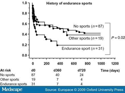
      Most of the described series include patients suffering concomitant AF and atrial flutter, suggesting that endurance sports contribute to the development of both arrhythmias. For example, Baldesberger et al.[27] found a higher incidence of flutter than AF in their series of veteran cyclists, although the authors did not describe whether these were common or atypical flutter episodes. Heidbuchel et al.[8] found that endurance athletes had a higher recurrence rate for AF than did controls (Figure 1). The authors conclude that endurance sport practice increases the risk of suffering AF after common flutter ablation. Hoogsteen et al.[29] found that 10% of athletes with AF also suffer episodes of atrial flutter. These observations suggest that both arrhythmias often co-exist in endurance athletes, and common flutter may be secondary to right atrial dilatation as a consequence of volume overload.

      Figure 1. 

      Patients with a history of endurance sports before ablation (n = 31) developed significantly more atrial fibrillation than controls or those with a history of other type o sports activity after flutter ablation (reproduced from reference 8, with permission).


      Atrial Fibrillation and Occupational Physical Activity

      These studies seem to have established that long-lasting endurance sport practice increases the risk of LAF. Vigorous physical activity associated with occupational activities may theoretically pose a similar risk. Data from the recently published GIRAFA study[10] appear to confirm this theory. The prospective GIRAFA study is conducted in consecutive patients with LAF recruited at the emergency room. In this case-control study, 107 LAF patients were compared with age- and sex-matched healthy controls. Total hours of physical activity (during work or leisure time) were collected with a detailed and validated questionnaire. For each physical activity, the following variables were recorded: age started, age ended, months per year, days per week, and hours per day. Subjects were asked to classify the intensity of each physical activity in four levels: sedentary, light, moderate, and heavy. The results showed that the moderate and heavy physical activity, whether sport- or job-related, increased the risk of suffering AF. In multivariable analysis, physical activity and atrial size were independent predictors for the development of AF, even after normalizing by body surface area (BSA) and height. In contrast with these observations, The Danish Diet Cancer and Health Study, conducted in a population of 19 593 men and 18 807 women with a mean age of 56 (range 50-65), failed to demonstrate any association between physical activities during working hours and risk of hospitalization with a diagnosis of AF or flutter.[30] This discrepancy may be due to the limited categorization and quantification of physical activity, compared to the much deeper analysis in the GIRAFA study. Further epidemiological studies, with a detailed quantification of work-associated physical activity, are needed to clarify this potential association.

      It is interesting to note the GIRAFA study's association of height and atrial size (absolute and normalized) with AF. In understanding the male predominance observed in AF, sex may indeed be secondary to that association. Hanna et al.[31] had already reported a relationship between stature and AF prevalence in patients with left ventricular (LV) dysfunction (Figure 2).

      Figure 2. 

      Thin-plate smoothing spline regression results assessing the non-linear relation between height and occurrence of lone atrial fibrillation (left panel), and the linear relation between left atrial (LA) diameter and occurrence of lone atrial fibrillation (right panel) (reproduced from reference 9, with permission).


      Pathophysiology of Sport-related Atrial Fibrillation

      What is the possible link between physical activity and AF? Several mechanisms may be acting together. It is well accepted that arrhythmias depend on triggers, substrates, and modulators, and these factors may be present in relation to physical activity (Figure 3).

      Figure 3. 

      Classical triangle of Coumel suggesting possible etiopathogenic factors influencing the development of atrial fibrillation in athletes.


      Triggers: Role of Atrial Ectopy

      Atrial ectopy, particularly pulmonary vein ectopy, has been shown to be the trigger in most episodes of paroxysmal AF.[32] Atrial and ventricular ectopy may be increased as a consequence of physical activity.[27,33] Moreover, increased ventricular ectopy in elite athletes is reversible after detraining.[34] Therefore, increased ectopy may be one of the mechanisms explaining the increased risk for AF associated with sport practice, provided that this ectopy acts upon an appropriate substrate. However, a recent paper by Baldesberger et al.[27] did not find an increased incidence of atrial ectopy, despite increases in ventricular ectopy and VT runs in former professional cyclists. Therefore, the hypothesis of increased atrial ectopy as an explanation for the association between sports and AF cannot be adequately sustained with currently available data.

      Modulators: Influence of Autonomic Nervous System

      Why does an apparently healthy individual start suffering from AF? Coumel[35] studied the influence of autonomic innervations in the appearance of AF and atrial flutter. He reported: 'Vagally mediated AF occurs more frequently in men than in women, with a ratio of ~4:1'. The age at which the first symptoms appear is classically between 40 and 50 years [sic]. The essential feature is the occurrence of the AF at night, often ending in the morning. Rest, the postprandial state (particularly after dinner) and alcohol are also precipitating factors [sic][36] (Figure 4). The author concluded that although AF occurred in a vagal context, an unidentified substrate probably existed. However, he did not establish a relationship between these episodes of AF and sport practice.

      Figure 4. 

      Twenty-four hours recording of heart rate showing a nocturnal episode of atrial fibrillation.


      Experimental data show that increased vagal tone shortens and increases the dispersion of the atrial refractory period, creating the conditions for re-entry.[37-39] However, vagal AF is considered to be a rare presentation of AF. This is probably due to the lack of systematic inquiry with patients. According to the GIRAFA study,[10] vagal AF is the rule rather than the exception in LAF patients (~70% of consecutive LAF patients had vagal AF). Therefore, the increased vagal tone induced by endurance sport practice may indeed facilitate the appearance of AF. In fact, heart rate is still lower in former athletes many years after cessation of professional training than in controls, as recently shown by Baldesberger et al.[27]

      Another interesting hypothesis recently raised by Swanson[40] in a review of the existing literature is that gastroesophageal reflux, which indeed has been proven to produce AF and vagal reflexes, may be the link between increased AF and exercise. However, this hypothesis has not yet been properly investigated.

      Arrhythmia Substrate

      Whether there is a structural substrate in LAF is still a matter of debate. In patients with hypertension or structural heart disease, it seems that AF is the consequence of structural changes in the atria (dilatation and fibrosis) secondary to chronic volume and pressure overload. It is therefore plausible that long-term endurance sport practice or occupational physical activity may induce structural changes in the atrium (enlargement, fibrosis) that may create a favourable substrate for the disease. In fact, Frustaci et al.[41] found structural changes in a series of 12 patients with paroxysmal, recurrent, drug refractory LAF. The authors described inflammatory lymphonomonuclear infiltrates, compatible with myocarditis, in 66% of patients; a non-inflammatory cardiomyopathic process in 17%; and patchy fibrosis in the remaining 17%. Whether the data correspond to a highly selected population cannot be definitively ruled out, but 100% of patients showed histological changes. On the other hand, these changes could have been produced by repetitive episodes of AF.

      A recent review of the literature by Swanson[42] shows that excessive endurance exercise and overtraining can lead to chronic systemic inflammation and there is a relationship between AF and C-reactive protein. Anti-inflammatory agents have been reported to lower C-reactive protein and ameliorate AF. Whether inflammation may be mediated by the renin-angiotensin system and a sustained increase in catecholamines remains to be elucidated. At present, no published studies combine these three concepts: AF, inflammation, and exercise. Additional studies are needed.

      Although the underlying mechanism for structural changes is not clear, recent echocardiographic data suggest that structural remodelling is often present in the atrium of elite athletes without AF. Pelliccia et al.[28] recently published a study that describes the remodelling induced by exercise in elite sport athletes. Their data showed that those involved in regular endurance practice have a larger atrium when compared with sedentary controls. Furthermore, a significant proportion (20%) showed enlarged atria according to established normal values.

      GIRAFA study data[10] showing that patients with LAF had a larger atrium when compared with controls suggest that subtle structural changes at the atrial level may account for the appearance of AF. The study further showed that patients with a first episode of AF had the same atrial size when compared with those suffering recurrences. Therefore, it seems that structural changes were present before onset of AF. On the other hand, patients with AF had larger LV mass, even after normalizing for BSA. This further supports the idea that exercise also had some repercussions in the ventricles, but without differences in diastolic function index when compared with controls. Although diastolic dysfunction has been proposed as the mechanistic background for atrial enlargement, it seems that volume and pressure overload act directly in the atrium, even before acting at the ventricular level.

      A recent case-control study by Lindsay and Dunn[43] involving 45 veteran athletes showed biochemical evidence of a disruption of the collagen equilibrium that would favour fibrosis. Athletes showed an increase in three collagen markers, plasma PICP, CITP, and TIMP-1, when compared with sedentary controls. The authors suggest that fibrosis occurs as part of the hypertrophic process in veteran athletes. Furthermore, an increase in fibrosis at the atrial and right ventricular level has been shown in a model of endurance exercise in rats.[44]

      Another factor that has been suggested as a cause of AF is the use of anabolic steroids. Although some isolated case reports show a link between AF and steroids,[45,46] the cases have presented in young athletes, at the moment of maximal physical activity, whereas AF in endurance sports seems to occur in middle-aged men, years after cessation of professional competitive or maximal activity. Therefore, although anabolic steroids may have a role in the genesis of AF, it is probably marginal. If atrial enlargement and fibrotic changes precede AF, what is the role of vagal tone and pulmonary veins in premature beats? It could be that in AF secondary to physical activity, vagal tone and ectopics may act more as a trigger and modulator than as the cause itself.

      Clinical Characteristics of Sport-related Atrial Fibrillation

      The typical clinical profile of sport-related AF or atrial flutter is a middle-aged man (in his forties or fifties) who has been involved in regular endurance sport practice since his youth (soccer, cycling, jogging, and swimming), and is still active. This physical activity is his favourite leisure time activity and he is psychologically very dependent on it. The AF is usually paroxysmal with crisis, initially very occasional and self limited, and progressively increasing in duration. Characteristically, AF episodes occur at night or after meals. As many as 70% of patients may suffer predominantly vagal AF.[10] They almost never occur during exercise. This makes the patient reluctant to accept a relationship between the arrhythmia and sport practice, particularly since his physical condition is usually very good. The crises typically become more frequent and prolonged over the years and AF becomes persistent. Progression to permanent AF has been described by Hoogsteen et al. in 17% of individuals in an observational series. In the GIRAFA study, 43% presented persistent AF.[10,29] The AF crisis frequently coexists with common atrial flutter in many patients, as previously discussed.

      Therapeutic Measures

      Although data on the reversibility of arrhythmia upon sport cessation are scarce, Furlanello et al.[25] have described a good response to sport abstinence in top-level athletes with AF. Our observations, although not systematic, suggest that limiting physical activity seems to significantly reduce the number of crises, particularly in those with recent onset and minimally dilated atrium. However, these patients are very dependent on physical activity and it is difficult for them to follow this advice. Previous studies have demonstrated the reversibility of hypertrophic changes at the ventricular level in the hearts of athletes. Biffi et al.[34] also showed a very significant decrease in ventricular ectopy upon sport cessation. Therefore, while awaiting more definitive data, it seems advisable to significantly reduce endurance sport practice in these cases.

      The possible long-term role of drugs (ACE inhibitors, angiotensin inhibitors, or beta-blockers) in preventing cardiac hypertrophy remains to be elucidated, although angiotensin blockers do seem to play a role in improving the results of cardioversion or AF ablation.[47,48] In terms of arrhythmia prevention, patients with recurrent episodes have been treated with flecainide and diltiazem, preventing 1:1 atrial flutter secondary to flecainide with good results. Some of them had undergone AF ablation with a success rate similar to patients not involved in endurance sport practice (authors' unplublished observations). In patients with predominant atrial flutter, ablation of the flutter is frequently associated with a higher incidence of AF recurrences, as pointed out by Heidbuchel et al.[8] A recent study by Furlanello et al.[49] described a highly successful ablation, with 90% success after a mean of two ablation procedures in a series of 20 athletes, without major complications. Apparently, the goal of the ablation was to allow rather veteran athletes (44 ± 13 years) to re-initiate their competitive activity. The reported series may represent a selected series of patients, since most of them presented exercise-induced AF, in contrast with the reported prevalence of vagal AF among endurance athletes. Although ablation seems to be quite effective, endurance sport cessation associated with drug therapy seems to us a more suitable approach as an initial therapy, particularly in non-professional, veteran athletes.

      Conclusions

      Vigorous physical activity, whether related to long-term endurance sport practice or to occupational activities, seems to increase the risk for recurrent AF. The underlying mechanisms remain to be elucidated, although structural atrial changes (dilatation and fibrosis) are probably present. There is a relationship between accumulated hours of practice and AF risk. Further studies are needed to clarify whether a threshold limit for the intensity and duration of physical activity may prevent AF, without limiting the cardiovascular benefits of exercise.


      Table 1. Summary of the Published Studies Analyzing the Relationship Between Atrial Fibrillation and Atrial Flutter and Endurance Sport Practice


      Studies Type of study Men (%) Age Type of sport(s) Cases/controls Odds ratio (CI) for AF in athletes
      Karjalainen et al.[5] Longitudinal case/control 100 47 ± 5 runners, 49 ± 5 controls Orienteers 262/373 5.5 (1.3-24.4)
      Mont et al.[6] Retrospective compared to general population 100 44 ± 13 athletes, 49 ± 11 non-athletes Endurance sports >3 h per week 70 lone AF 61% in male athletes with lone AF
      Elosua et al.[7] Retrospective case/control 100 41 ± 13 AF pat, 44±11 controls Endurance sports: current practice and >1500 accumulated hours of practice 51/109 2.87 (1.39-7.05) adjusted for age and hypertension
      Heidbuchel et al.[8] Case/control in patients undergoing flutter ablation 83 53 ± 9 sports, 60 ± 10 controls Cycling, running, or swimming >3 h per week 31/106 1.81 (1.10-2.98)
      Molina et al.[9] Longitudinal case/control 100 39 ± 9 runners, 50 ± 13 sedentary Marathon runners 252/305 8.80 (1.26-61.29) adjusted for age and blood pressure
      Baldesberger et al.[27] Longitudinal case/control 100 67 ± 7 cyclist, 66 ± 6 golfers Cyclists 134/62 10% AF in cyclists, 0% AF in controls
      Mont et al.[10], GIRAFA study Prospective case/control 69 48 ± 11 Endurance sports 107/107 7.31 (2.33-22.9), >550 h of accumulated heavy physical activity


        References

        1. Morris JN, Everitt MG, Pollard R, Chave SP, Semmence AM. Vigorous exercise in leisure-time: protection against coronary heart disease. Lancet (1980) 2:1207-10.
        2. Kujala UM, Kaprio J, Taimela S, Sarna S. Prevalence of diabetes, hypertension, and ischemic heart disease in former elite athletes. Metabolism (1994) 43:1255-60.
        3. Blair SN, Kampert JB, Kohl HW 3rd, Barlow CE, Macera CA, Paffenbarger RS Jr, et al. Influences of cardiorespiratory fitness and other precursors on cardiovascular disease and all-cause mortality in men and women. JAMA (1996) 276:205-10.
        4. Thompson PD, Buchner D, Pina IL, Balady GJ, Williams MA, Marcus BH, et al. Exercise and physical activity in the prevention and treatment of atherosclerotic cardiovascular disease: a statement from the Council on Clinical Cardiology (Subcommittee on Exercise, Rehabilitation, and Prevention) and the Council on Nutrition, Physical Activity, and Metabolism (Subcommittee on Physical Activity). Circulation (2003) 107:3109-16.
        5. Karjalainen J, Kujala UM, Kaprio J, Sarna S, Viitasalo M. Lone atrial fibrillation in vigorously exercising middle aged men: case-control study. BMJ (1998) 316:1784-5.
        6. Mont L, Sambola A, Brugada J, Vacca M, Marrugat J, Elosua R, et al. Long-lasting sport practice and lone atrial fibrillation. Eur Heart J (2002) 23:477-82.
        7. Elosua R, Arquer A, Mont L, Sambola A, Molina L, Garcia-Moran E, et al. Sport practice and the risk of lone atrial fibrillation: a case-control study. Int J Cardiol (2006) 108:332-7.
        8. Heidbuchel H, Anne W, Willems R, Adriaenssens B, Van de WF, Ector H. Endurance sports is a risk factor for atrial fibrillation after ablation for atrial flutter. Int J Cardiol (2006) 107:67-72.
        9. Molina L, Mont L, Marrugat J, Berruezo A, Brugada J, Bruguera J, et al. Long-term endurance sport practice increases the incidence of lone atrial fibrillation in men: a follow-up study. Europace (2008) 10:618-23.
        10. Mont L, Tamborero D, Elosua R, Molina I, Coll-Vinent B, Sitges M, et al. Physical activity, height, and left atrial size are independent risk factors for lone atrial fibrillation in middle-aged healthy individuals. Europace (2008) 10:15-20.
        11. Taggar JS, Lip GY. Risk predictors for lone atrial fibrillation. Europace (2008) 10:6-8.
        12. Lampert R. Atrial fibrillation in athletes: toward more effective therapy and better understanding. J Cardiovasc Electrophysiol (2008) 19:463-5.
        13. Schoonderwoerd BA, Smit MD, Pen L, Van Gelder I. New risk factors for atrial fibrillation: causes of 'not-so-lone atrial fibrillation. Europace (2008) 10:668-73.
        14. Mozaffarian D, Furberg CD, Psaty BM, Siscovick D. Physical activity and incidence of atrial fibrillation in older adults. The Cardiovascular Health Study. Circulation (2008) 118:800-7.
        15. Fuster V, Ryden LE, Cannom DS, Crijns HJ, Curtis AB, Ellenbogen KA, et al. ACC/AHA/ESC 2006 guidelines for the management of patients with atrial fibrillation-executive summary: a report of the American College of Cardiology/American Heart Association Task Force on Practice Guidelines and the European Society of Cardiology Committee for Practice Guidelines (Writing Committee to Revise the 2001 Guidelines for the Management of Patients With Atrial Fibrillation). J Am Coll Cardiol (2006) 48:854-906.
        16. Benjamin EJ, Wolf PA, D'Agostino RB, Silbershatz H, Kannel WB, Levy D. Impact of atrial fibrillation on the risk of death: the Framingham Heart Study. Circulation (1998) 98:946-52.
        17. Wolf PA, Benjamin EJ, Belanger AJ, Kannel WB, Levy D, D'Agostino RB. Secular trends in the prevalence of atrial fibrillation: the Framingham Study. Am Heart J (1996) 131:790-5.
        18. Wang TJ, Parise H, Levy D, D'Agostino RB Sr, Wolf PA, Vasan RS, et al. Obesity and the risk of new-onset atrial fibrillation. JAMA (2004) 292:2471-7.
        19. Brugada R, Tapscott T, Czernuszewicz GZ, Marian AJ, Iglesias A, Mont L, et al. Identification of a genetic locus for familial atrial fibrillation. N Engl J Med (1997) 336:905-11.
        20. Leather RA, Kerr CR. Atrial fibrillation in the absence of overt cardiac disease. In: Atrial fibrillation: Mechanisms and Management. (1992) New York: Raven Press. 93-108.
        21. Planas F, Romero-Menor C, Vazquez-Oliva G, Poblet T, Navarro-Lopez F. Natural history of and risk factors for idiopathic atrial fibrillation recurrence (FAP Registry). Rev Esp Cardiol (2006) 59:1106-12.
        22. Levy S, Maarek M, Coumel P, Guize L, Lekieffre J, Medvedowsky JL, et al. Characterization of different subsets of atrial fibrillation in general practice in France: the ALFA study. The College of French Cardiologists. Circulation (1999) 99:3028-35.
        23. Coumel P. Paroxysmal atrial fibrillation: a disorder of autonomic tone? Eur Heart J (1994) 15:9-16.
        24. Coelho A, Palileo E, Ashley W, Swiryn S, Petropoulos AT, Welch WJ, et al. Tachyarrhythmias in young athletes. J Am Coll Cardiol (1986) 7:237-43.
        25. Furlanello F, Bertoldi A, Dallago M, Galassi A, Fernando F, Biffi A, et al. Atrial fibrillation in elite athletes. J Cardiovasc Electrophysiol (1998) 9:S63-8.
        26. Masia R, Pena A, Marrugat J, Sala J, Vila J, Pavesi M, et al. High prevalence of cardiovascular risk factors in Gerona, Spain, a province with low myocardial infarction incidence. REGICOR Investigators. J Epidemiol Commun Health (1998) 52:707-15.
        27. Baldesberger S, Bauersfeld U, Candinas R, Seifert B, Zuber M, Ritter M, et al. Sinus node disease and arrhythmias in the long-term follow-up of former professional cyclists. Eur Heart J (2008) 29:71-8.
        28. Pelliccia A, Maron BJ, Di Paolo FM, Biffi A, Quattrini FM, Pisicchio C, et al. Prevalence and clinical significance of left atrial remodeling in competitive athletes. J Am Coll Cardiol (2005) 46:690-6.
        29. Hoogsteen J, Schep G, Van Hemel NM, Van Der Wall EE. Paroxysmal atrial fibrillation in male endurance athletes. A 9-year follow up. Europace (2004) 6:222-8.
        30. Frost L, Frost P, Vestergaard P. Work related physical activity and risk of a hospital discharge diagnosis of atrial fibrillation or flutter: the Danish Diet, Cancer, and Health Study. Occup Environ Med (2005) 62:49-53.
        31. Hanna IR, Heeke B, Bush H, Brosius L, King-Hageman D, Beshai JF, et al. The relationship between stature and the prevalence of atrial fibrillation in patients with left ventricular dysfunction. J Am Coll Cardiol (2006) 47:1683-8.
        32. Haissaguerre M, Jais P, Shah DC, Takahashi A, Hocini M, Quiniou G, et al. Spontaneous initiation of atrial fibrillation by ectopic beats originating in the pulmonary veins. N Engl J Med (1998) 339:659-66.
        33. Bjornstad H, Storstein L, Meen HD, Hals O. Ambulatory electrocardiographic findings in top athletes, athletic students and control subjects. Cardiology (1994) 84:42-50.
        34. Biffi A, Maron BJ, Verdile L, Fernando F, Spataro A, Marcello G, et al. Impact of physical deconditioning on ventricular tachyarrhythmias in trained athletes. J Am Coll Cardiol (2004) 44:1053-8.
        35. Coumel P. Paroxysmal atrial fibrillation: a disorder of autonomic tone? Eur Heart J (1994) 15:9-16.
        36. Coumel Ph. Neural aspects of paroxysmal atrial fibrillation. In: Atrial Fibrillation: Mechanisms and Management—Falk RG, Podrid PhJ, eds. (1992) Raven Press. 109-25.
        37. Hoff HE, Geddes LA. Cholinergic factor in auricular fibrillation. J Appl Physiol (1955) 8:177-92.
        38. Moe GK, Abildskov JA. Atrial fibrillation as a self-sustaining arrhythmia independent of focal discharge. Am Heart J (1959) 58:59-70.
        39. Alessi R, Nusynowitz M, Abildskov JA, Moe GK. Nonuniform distribution of vagal effects on the atrial refractory period. Am J Physiol (1958) 194:406-10.
        40. Swanson DR. Running, esophageal acid reflux, and atrial fibrillation: a chain of events linked by evidence from separate medical literatures. Med Hypotheses (2008) 71:178-85.
        41. Frustaci A, Chimenti C, Bellocci F, Morgante E, Russo MA, Maseri A. Histological substrate of atrial biopsies in patients with lone atrial fibrillation. Circulation (1997) 96:1180-4.
        42. Swanson DR. Atrial fibrillation in athletes: implicit literature-based connections suggest that overtraining and subsequent inflammation may be a contributory mechanism. Med Hypotheses (2006) 66:1085-92.
        43. Lindsay MM, Dunn FG. Biochemical evidence of myocardial fibrosis in veteran endurance athletes. Br J Sports Med (2007) 41:447-52.
        44. Benito B, Gay-Jordi G, Serrano A, Sirenko V, Tamborero D, Berruezo A, et al. Chronic exercise induces atrial and right ventricular fibrosis in a rat model. (Abstract Supplement). Eur Heart J (2008) 29:740.
        45. Sullivan ML, Martinez CM, Gallagher EJ. Atrial fibrillation and anabolic steroids. J Emerg Med (1999) 17:851-7.
        46. Lau DH, Stiles MK, John B, Shashidhar, Young GD, Sanders P. Atrial fibrillation and anabolic steroid abuse. Int J Cardiol (2007) 117:e86-e87.
        47. Anne W, Willems R, Van der MN, Van de WF, Ector H, Heidbuchel H. Atrial fibrillation after radiofrequency ablation of atrial flutter: preventive effect of angiotensin converting enzyme inhibitors, angiotensin II receptor blockers, and diuretics. Heart (2004) 90:1025-30.
        48. Madrid AH, Bueno MG, Rebollo JM, Marin I, Pena G, Bernal E, et al. Use of irbesartan to maintain sinus rhythm in patients with long-lasting persistent atrial fibrillation: a prospective and randomized study. Circulation (2002) 106:331-6.
        49. Furlanello F, Lupo P, Pittalis M, Foresti S, Vitali-Serdoz L, Francia P, et al. Radiofrequency catheter ablation of atrial fibrillation in athletes referred for disabling symptoms preventing usual training schedule and sport competition. J Cardiovasc Electrophysiol (2008) 19:457-62.
        Acknowledgements

        We thank Elaine M. Lilly, PhD, Writer's First Aid and Neus Portella, Research Assistant for editing the mauscript.